Enhance your health with free online physiotherapy exercise lessons and videos about various disease and health condition
CERVICAL RIB SYNDROME
Cervical Rib refers to an abnormal protrussion in the cervical region which can either be due to abnormal enlargement of the transverse process of C7 vertebra or a small rib or fibrous band running from the 7th cervical vertebra to the first true rib or to the sternum but usually it is present posteriorly up-to a short distance.
It is usually diagnosed in middle age group persons though is present since birth. The cause is that by middle age, the shoulders start drooping which causes the cervical rib to get depressed and hence compressing the nerve root of the concerned region. This rib is usually asymptomatic but it may give rise to neurological symptoms if it exerts pressure on the subclavian artery or the brachial plexus like-
- paraesthesia of hand
- hypothenar wasting
- atonia in the muscles of the shoulder girdle.
A cervical rib is a supernumerary (extra) rib which arises from the seventh cervical vertebra. It is a congenital abnormality located above the normal first rib. This rib is present in only about 1 in 200 (0.5%) of people; in even rarer cases, an individual may have not one but two cervical ribs. The presence of a cervical rib can cause a form of thoracic outlet syndrome due to compression of the brachial plexus or subclavian artery. Compression of the brachial plexus may be identified by weakness of the muscles around the muscles in the hand, near the base of the thumb. Compression of the subclavian artery is often diagnosed by finding a positive Adson's sign on examination, where the radial pulse in the arm is lost during abduction and external rotation of the shoulder.
 |
 |
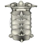 |
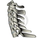
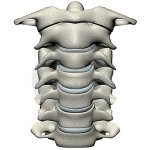
| human-lateral-cervical-spine | human-anterior-cervical-spine | human-posterior-cervical-spine |
Cervical Rib History
- 150 AD: Galen, the Greek anatomist and court physician to Marcus Aurelius, first describes the Cx rib in human dissections.
- 1500’s: Andreas Vesalius, who is responsible for the revival of Galen’s work (which had fallen into disfavor in the Dark Ages), again describes the Cx rib in human cadavers.
- 1742: Francois-Joseph Hunauld describes and categorizes supernumerary ribs in humans, including the Cx rib.
- 1821: Sir Astley Cooper first describes arterial thoracic outlet syndrome in a young woman with arm ischemia, and demonstrates the relationship between a Cx rib andcompression of the subclavian artery.
- 1831: Henry Mayo in London reports the first subclavian artery aneurysm due to thoracic outlet syndrome, caused by an exostosis of the first rib.
- 1853: John Hilton, a British surgeon, describes gangrene of the arm in a patient with compression of the subclavian artery caused by an exostosis of the first rib.
- 1860: W. H. Willshire notes the relationship between a Cx rib and paresthesias of the upper extremity.
- 1861: Richard Holmes Coote at St. Bartholomew’s Hospital in London performs the first surgical resection of a cervical rib, with relief of arterial TOS.
- 1903: F. Bramwell describes neurovascular compression in the presence of a normal first rib, without a Cx rib.
- 1905: John Benjamin Murphy of Chicago was the first surgeon to resect a cervical rib that was associated with a subclavian artery aneurysm.
- 1907: Dr. William Keen at Thomas Jefferson University in Philadelphia publishes a review of 42 cases of resected cervical ribs, and describes a clinical definition and surgical treatment for this disorder.
- 1931: Telford and Stopford propose that the variable distribution of sympathetic fibers in the brachial plexus could explain the variable degrees of vascular disturbance seen inpatients with symptomatic cervical ribs.
- 1943: Murray Falconer and L.G. Weddell describe several military recruits with neurovascular compression caused by heavy backpacks and assuming the "military" position, with the shoulders thrust backwards. They believe that this neurovascular compression is caused by compression of the neurovascular bundle between the normal clavicle and first rib on assumption of an unusual posture, even in the presence of normal anatomy, and coined the term “costoclavicular syndrome”. Dr. Falconer publishes a second paper with Dr. Franklin W. P. Li, describing an additional 11 patients who underwent first rib resection with good results. Several of these patients had been previously treated for carpal tunnel syndrome, without relief.
Clinical Features
Local Symptoms
There is often a tender supraclavicular lump which is bony hard and is fixed when palpated.
Sensory Symptoms
- (a) Tingling in hands or fingers; confined either to radial side or ulnar side or sometimes involve even whole hand.
- (b) Pain may sometimes radiate downwards from the arm.
Motor Symptoms
- (a) Loss of gripping power of the hand.
- (b) Tendency of dropping things from the hand.
- (c) Wasting of palmar muscles; either thenar or hypothenar or interossei muscles.
Vascular Symptoms
- (a) Cold and clumsy extremities, particularly the fingers.
- (b) Skin colour changes to blue associated with trophic changes.
- (c) There is rare risk of gangrene.
- (d) Radial pulse becomes feeble or may even be absent.
Diagnosis
1- Mainly by X-ray to detect presence of Cervical ribs , which could be easily palpated.
2. Special Tests
Adson's Test
- Indications- Evaluation of Cervical Ribs/ Thoracic Outlet Syndrome.
- Technique- Patient breathes deeply, Neck extended, Chin turned toward affected side. The examiner lifts the arm away to the side to 90 degrees and performs external rotation of the shoulder, and notes whether the radial pulse disappears. However there are many false positives, as the radial pulse may disappear in normal people as the head of the humerus (upper arm bone) compresses the brachial vessels when the arm is taken beyond 90 degrees. Repeat test with chin to opposite side.
- Interpretation- Positive test finding ( Decreased Radial Pulse and/or Distal extremity pain reproduced ) suggests interscalene compression.
Foraminal Compression Test/ Spurling's Test
Spurling's test is an orthopedic test used to diagnose nerve root compression primarily at the cervical level. It should not be used in instances in which vertebral instability is suspected.
Shoulder Abduction Test
Shoulder Abduction Test is an orthopedic test used to help diagnose a cervical nerve root injury or cervical disc herniation. It is performed by having the patient abduct their shoulder and place their hand on top of their head. A positive test will involve a decrease in radiculopathy or pain.
Differential Diagnosis
The differential diagnosis for Cervical Ribs is quite broad and includes neurologic, vascular, pulmonary, cardiac, and esophageal disorders.
- 1)Some of the more common conditions include herniated cervical disk, cervical spondylosis, and peripheral neuropathies.
- 2) Peripheral vascular disease like Raynaud's disease.
- 3) Neurological conditions-like syringomyelia, polio, muscular dystrophy, motor neuron disease.
Medical treatment
Anti-inflammatory drugs and analgesics are provided as a conservative means of treatment.
Surgical treatment
surgery is essential in conditions of severe, progressive vascular and neurological signs and symptoms which are unbearable for the patients. It includes:
(a) Removal of extra segment.
(b) Dividing the scalene group of muscles.
Physiotherapy Treatment
Read Research Articles about Cx Rib on PubMed
On the basis of symptoms of the patient, the regime of physiotherapy is planned.
- For pain relief- Heat modalities are used like short wave diathermy but it should not be applied in case of sensory impairments.
- To improve distal circulation- Exercises of hand and finger should be started.
- TO improve tone, power and endurance- Strengthening exercises of whole arm perticularly small muscles of the arm.
- Correction of posture by postural guidance- In this, patient is taught to use postural mirror to see that his shoulders are in level, head is straight, looking forward.
- Planning specific exercises- To develop particular muscles groups for specific movements of shoulder girdle like elevation, retraction, and raising the arm overhead as these movements brings spontaneous relief. The important exercises are-
1-Self resisted scapular elevation.
2-Self resisted scapular adduction.
3-Endurance training exercise for the shoulder girdle muscles.
4-Progressive resistance exercises for shoulder girdle muscles with weight. - Deep Tissue Massage for TOS ( thoracic Outlet Syndrome).
Return from Cervical Rib to Home Page
Recent Articles
|
Author's Pick
Rating: 4.4 Votes: 252 |


 About Prodyut Das (PT, DPT) Physical Therapist at SMC, New York, USA. Former PT Winner Regional Health, South Dakota, Former HOD Physiotherapy & Fitness center @ NIMT Hospital, Greater Noida. Former PT ISIC Hospital. DPT ( Univ of Montana), MPT (neuro), MIAP, cert. manual therapist, Medical Neuroscience (USA). Licensed Physical Therapist in NY and Texas, USA. |
Connect with me- |
© Copyright physiotherapy-treatment.com since 2009 |