Enhance your health with free online physiotherapy exercise lessons and videos about various disease and health condition
Extracorporeal Shock Waves in Orthopaedics
Contents
In 1980, the first patient with renal calculi was successfully treated with minimally invasive extracorporeal shock waves lithotripsy (ESWL). Since then the medical field of application of this form of energy has been extended. The most recent result of the medical and technical development of application is extracorporeal shock wave therapy (ESWT) for the treatment of orthopaedic pathologies.
Valchanov and Michailov and Sukul et al. inaugurated ESWT for the treatment of delayed and non-union of fractures, describing local decortication and fragmentation with stimulation of osteogenesis. These positive aspects were corroborated by Haist et al., who noticed bony consolidation in 32 out of 40 cases with a pseudoarthrosis. In case reports, Dahmen et al. and Loew and Jurgowski achieved good results in calcifying tendinitis of the shoulder. The indications for ESWT are progressively moving to cover a large part of the rest of the tendinous pathologies, especially at the insertional areas, and their limits of application, on the bone side of the orthopaedic diseases, have already reached the field of osteochondritis and osteonecrosis of the hip and the knee as a new promising technique of treatment.
Definition
A shock wave is scientifically defined as an acoustic or sonic wave, at the wave front of which the pressure above atmospheric rises from ambient pressure to maximum pressure (amplitude) within a few nanoseconds. Current therapeutically used pressure amplitudes, range between 10MPa and more than 100MPa (I MPa = 10 bar, approximately tenfold atmospheric pressure). As a shock wave propagates at a specific speed - as do all sonic waves - determined by the medium in which it propagates and the intensity of the shock wave itself, one can calculate the shock front thickness. This is the spatial dimension between the locations where the pressure amplitude has been reached.
Sources
In practice, all extracorporeal shock waves (ESW) generators consist of an electrical energy source, an electroacoustic conversion mechanism and a device for focusing the sound waves.
There are three methods of generating shock waves, namely by piezoelectic, electrohydraulic, or electromagnetic means.
Without entering into a detailed discussion on the technical data about the Extracorporeal Shock Waves generators, it is important to know that the electrohydraulic system produces high energy Extracorporeal Shock Waves (ESW), but with marked oscillations in the values of the energy flux density through the tissues, which do not remain constant during the therapeutic application. They need a spark electrode to work, but a fast deterioration in use reduces its life to about 1000 sparks before the electrode needs to be changed, while a single application, lasts some thousands of impulses, on average. The piezoelectric device is very precise and has a long life, but, in spite of a very sophisticated technology, produces only low-energy Extracorporeal Shock Waves (ESW), often requiring, repeated treatments to reach the desired result.
The more actual electromagnetic generators are mainly differentiated into two types because of their shapes: flat and cylinder coils. The average value of pressure, expressed in terms of energy flux density, is almost constant and the coil is a very long-lasting device. To achieve a specific effect, the sound waves must be focused. When employing point sources (e.g., sparks) or cylinder sources, a focusing mirror is used. When employing areal sources, lenses or a self focusing appliance of partially spherical shape are used.
Extracorporeal Shock Waves in Medical Practice
Shock waves employed in medical practice are characterized by high positive press ures up to 80 - 100 MPa and negative pressures of 5 - 10 MPa Furthermore, they have a short rise time of 30 - 120 ns and a shorter pulse duration (5 μs),. In contrast to ultrasound, shock waves have low frequencies. Just in this respect, there is less absorption by the tissue. Moreover, the shock waves are applied with a lower repetition frequency of 1- 2, maximum 4 Hz, which means that they have a low time-averaged intensity. The only thing that can be said for sure is that the shock wave does not cause tissue warming. None of the known shock wave effects are due to thermal effects.
During the ESWT, it is important to know the energy dose administered for each treatment. All the authors, under a general agreement, distinguish the Extracorporeal Shock Waves into high-energywaves and low-energywaves; the level of energy turns out to be a complex parameter which is defined by the ratio between the applied power and the tissue density.
Treatments with intermediate and high energy levels produce energy flux densities which range from those used for treatment of nephrolithiasis to many times this energy level, i.e., densities from 0.37 to 1.2 mJ/mm2• This form of treatment nearly always requires analgesia or anesthesia. The indications are represented by pseudoarthrosis and some species of tendinosis calcharea.
Low-energy treatment employes 10% - 20% of the dose used for renal calculi, i.e., densities between 0.08 and 0.23 mJ/mm2• In most cases anesthesia or analgesia is not required. The low energy range is mainly reserved for the treatment of localized soft tissue disorders.
Modes of Action
Regarding the action of shock waves on tissue, four phases have been postulated:
1. Physical phase: extracellular cavitations, ionized molecules and an increase of membrane permeability are direct effects of the shock waves.
2 . Physical-chemical phase: diffusible radicals and interactions with biomolecules (in both phases mitochondriallesions were observed).
3. Chemical phase: may be accompanied by intracellular reactions and molecular changes.
4. Biological phase: noted if these changes persist.
Many of the shock wave tissue interactions are not yet completely understood. Several theories have been formulated to explain the mechanism of action of Extracorporeal Shock Waves on bone-related soft tissue pain:
1. The shock wave locally alters the chemical environment in such a way, that pain-inhibiting substances are produced.
2. The shock wave damages the cell membrane; no more generator potential is produced in the pain receptor for the formation of the pain signals.
3. The shock wave stimulates the pain receptor to emit a high frequency of nerve impulses. The retransmission of pain impulses is inhibited as it is subject to external stimuli (gate-control theory).
4. The shock wave induces arelease of endorphin resulting in the reduction of (local) sensitivity to pain.
Therefore, there seem to be two possible modes of action in tissue; direct effects, or those caused by cavitation.
Direct Effects. Direct effects are mechanical effects caused by temporary positive pressure amplitudes during the propagation of a pressure pulse. The stability under load of the material (tissue) may be exceeded at the interface between the materials with different acoustic impedance. At such interfaces, the shock waves lead to changes in the excursion, which result in tensile and shearing loads. In soft tissue, where there are no great differences in acoustic impedance, the direct effects of shock waves do not appear to play a significant role.
Cavitation. The definition of cavitation is: expansion and oscillation of gas-filled cavities (pre-existing as cavitation nuclei) due to the tensile portion of a pressure wave with subsequent collapse. The cavitation results in mechanical and chemical effects:
1. Mechanical: shearing loads develop due to the oscillation of the developed cavitation bubbles. Due to the collapse of the bubbles, there is an inflow of water (jet streams). These inflowing water masses can reach velocities of between 400 and 700 m/s , which correspond to the velocity of bullets. One can surely imagine an effeet of this jet stream on a mechanical basis.
2. Chemical: during the collapse of the cavitation bubbles, high temperatures develop locally inside the bubbles, which lead to the development of radicals. The induced radicals can subsequently lead to damage in the tissue.
In experiments on cells in suspension, Brümmer and Smits found a cell damage pattern which is dependent on the number of shock waves and the shock wave energy applied:
1. Transient membrane rupture (permeability) with secondary damage (cell edema)
2. Transient damage to endoplasmatic reticulum and cell nucleus
3. Dose-dependent vacuolization of the cytoplasma
4. Lesion in cytoskeleton (actin and vimentin fibers)
5. Complete cell rupture (e.g., hemolysis)
To understand the mode of action of the Extracorporeal Shock Waves (ESW) on the bone tissue we need to consider their mechanical effects on the bone trabeculae. When the shock wave hits the anterior surface of a trabecula, because the impedance inside it (II medium) is lower compared with the one outside it (I medium), at the level of the I interface (I medium/II medium), part of the energy is reflected back (stretching force), while the remaining energy ( compressing force) advances through the trabecula (II medium) to reach the posterior surface of it up to the II interface (II medium/I medium), where the acoustic impedance gradient is inverted and the reflected wave is negative.
At the level of both interfaces, the cavitation bubbles are formed and are then submitted to a deforming compression by the gradient of pressure, with a consequent reduction of the volume and increase in energy; they collapse progressively, the anterior wall hit by the shock wave introflects, while the posterior wall is extroflected by the stretching components, until the bubble releases part of the stored energy generating the jet streams directed toward the gradual shattering of the trabeculae.
According to Sukul and Johannes these effects on the bone structures are comparable to the effects of lithotripsy on kidney and gallbladder stones. In the literature, several hypotheses have been considered:
1. Microfissures at the fracture ends
2. Development of a hematoma
3. Development of small cortical/spongiosa chips
4. Transformation of local connective-tissue cells
Also, the phenomenon of neoangiogenesis might be related to the presence of chemiotactic factors, very close to the ESAF (endothelial stimulating angiogenetic factor), released by the stress fibers.
Extracorporeal Shock Waves (ESW) has important effects on nerves and nerve cells. It could be hypothetically assumed that the mode of action is based on a direct mechanical effect of the shock wave on the axon membrane in the sense of an increase in permeability with consecutive depolarization and triggering of an action potential. However, it appears more probable that a cavitation-based effect plays a role in the triggering of the cumulative action potentials. A hyperpolarization of the neurons is achieved by means of shock wave treatment, whereby an energy dependency was discovered, i.e., the higher the energy of the emitted shock wave, the greater the polarization. These effects lasted for approximately 1 h after the shock wave. During hyperpolarization, a stimulus is necessary which is larger than that required before shock wave treatment, to excite an action potential. Whether this effect could play a role in palliative therapy will require further study.
Indications and Protocols of Therapy
From the first reports of Valchanou and Michailov who inaugurated high energy ESWT for the treatment of delayed and non-union fractures, the clinical indications have been largely expanded toward other orthopaedic disorders, both in hard tissue pathology, i.e., osteonecrosis and osteochondritis or the problems of osteointegration at the metal-bone interface of the loose cementless prosthesis, and the soft tissue pathologies, i.e., almost all the insertional tendinitis and enthesopathies or muscles strains.
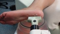
There is a general agreement among the authors that high energy treatments should be reserved for bone affections whereas low energy ESWT is the most indicated treatment for soft tissues lesions. During soft tissue therapies, usually without any form of analgesia or anesthesia, the application of shock waves is also based on a sort of 'pain feedback', by asking the patient when he recognizes his symptomatology, and the quality and the site of pain; but it must always be remembered that there are algogenic structures, i.e., the periosteum of the superficial bones or the sensitive nerve bundles, easily stimulated by the Extracorporeal Shock Waves (ESW): their activation always induces strong pain and it might confuse the operator. For this reason it is important that an in-line ecographic aiming device is available to precisely individuate the site of pathology.
The high energy ESWT almost always requires pain control by means of analgesic/anesthetic treatment. An image intensifier is absolutely necessary to position the focus of the Extracorporeal Shock Waves (ESW) right on the site of pathology, otherwise it is impossible to individuate it exactly.
Pseudarthrosis
Delayed union is defined when a fracture is not healed completely within 4 months. Healing that does not happen within 6months is called pseudarthrosis. Congenital forms of pseudarthrosis are known. According to radiological criteria, pseudoarthrosis can be hypertrophie or atrophie. The present studies show that ESWT is not indicated in cases of congenital non-unions, and that results are better in hypertrophie forms than in atrophie ones.
The protocol schemes according to Russo et al. distinguish between small bones and other bones. In the first case, two applications, the latter 24 - 48 h after, 4000 impulses, 0.7-1.0 mJ/mm2, are recommended.
Non-unions of the long bones are treated with Extracorporeal Shock Waves (ESW) in four sessions, each second day, 4000/5000 impulses, 1.0 mJ/mm2• The decision to cease the treatment or to continue it with the second phase is based on the evidence of osteogenic response in the X-rays and magnetic resonance imaging (MRI) controls. A second and a third phase are only needed when partial fusion or no fusion at all are observed. Modalities and energy rates are the same as for the first phase.
Prosthesis Loosening
During the last few years, the application of Extracorporeal Shock Waves (ESW) for hip prosthesis failure was advocated from two different, almost opposite, points of view. Haupt and other authors have considered the possibility of using it to facilitate the rem oval of cement at the interface with bone in the revision of cemented prostheses.
On the other hand, Schaden believes that the application of ESW for loosening of the femoral component in cementless hip prostheses might effectively help to re-fix the stern bone, as he observed at least in three case reports. What he advises is to utilize a high energy device (28kV) for a number of applications of between 4000 and 8000 impulses.
Osteochondritis, Osteochondrosis, and Osteonecrosis
Osteochondritis dissecans of the knee or talus was treated with Extracorporeal Shock Waves (ESW) by Schieberger, resulting in integration of the dissecating bone piece, but success rates have not been reported yet. Few treatments with promising results have been performed in patients with Köhler's, Perthes's, or Osgood-schlatter's disease. To date, these therapies must be considered experimental.
Some authors have started, in the last 2 or 3 years, to employ Extracorporeal Shock Waves (ESW) in the therapy of earlier forms of osteonecrosis of the femoral head and femoral condyles of the knee. Unfortunately, we have not yet had results or technical information about these procedures.
Tendinosis Calcarea of the Shoulder
Probably, calcifying tendinitis of the shoulder represents the first field of application of Extracorporeal Shock Waves (ESW) in orthopaedic soft tissue pathologies.
According to Rompe, high energy Extracorporeal Shock Waves (ESW) are expected to exert a direct, mechanically disintegrating effect on hard surfaces such as calcareous deposits in the supraspinous tendon. Low energy ESWT, however, is regarded as a form of hyperstimulation analgesia. In the low energy treatment the patient receives 1500 impulses of 0.06 mJ mm-2 of energy density. The high energy treatment consists of 1500 impulses of 0.28 mJ mm-2 administered under regional anesthesia.
Gigliotti, Russo et al. propose a different protocol of treatment for calcifying tendinitis: 2000 - 2500 impulses set at a level of density energy of 0.07 - 0.11 mJ mm-2 in a series of 5-weekly sessions.
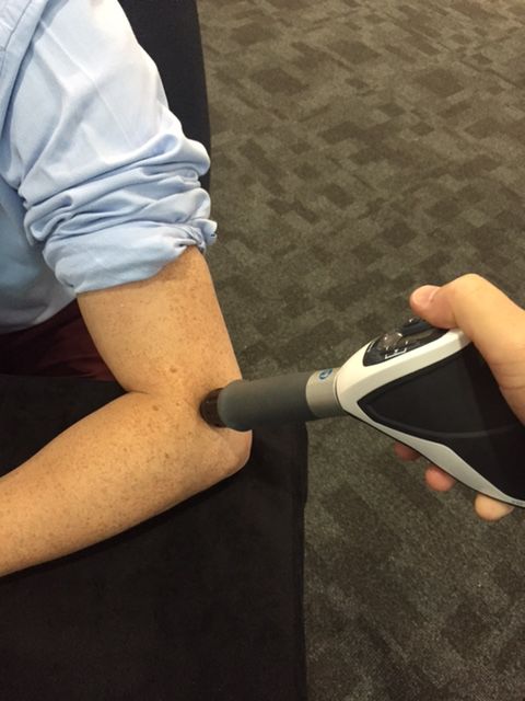
Insertional Tendinitis
Insertional tendinitis is a very common pathological picture in athletes. Among these, patellar tendinitis and epicondylitis humeri (radialis more often than ulnaris) have the higher rates of incidence. Haist [78] proposes a treatment protocol based on three to four sessions of about 25min (1500 impulses at a frequencyof 1Hz) with the energy density between 0.06 and 0.12 mJ mm-2• We had promising results in patellar tendinitis after submitting the patients to a four-session cyde of treatment: 1500 impulses at an energy level of 0.11 mJ mm-2 each seventh day. No patient needed analgesia for it.
Contraindications and Side Effects
ESWT is absolutely contraindicated in pediatric or adoIescent patients under the age of 18 years, during pregnancy, if there are pathological neurological, or vascular findings, if there are local infections, tumors, or blood coagulation diseases.
Rotator cuff tears, arthritic degenerative changes of the gleno-humeral joint, and ligamentous articular instability should also be considered relative contraindications for treatment.
When properly performed, ESWT is free from general side effects in healthy subjects.
Regional reactions have been observed since the first urological employment. They are usually temporary and limited to small local swelling, hematomas and petechial hemorrhages.
Conclusions
According to the opinion of the several authors involved up to now in ESWT, shock wave application in orthopaedics has to be considered as minimally invasive, and a safe method of treatment as regards traditionally difficult and problematic bone and tendinous pathologies, like non-unions and enthesopathies. Almost no side effects whatsoever have been observed in hundreds of thousands of therapy sessions all over the world since the very early beginnings of this treatment.
However, due to the high cost of the equipment, and consequently of the treatments, this therapy should be restricted to patients who do not experience considerable pain relief after a defined period of established conventional treatment.
When non-union of a fracture occurs, either in operated and non-operated patients, ESWT should be considered, and eventually administered, before resorting to a surgical treatment, as a revision or completely new procedure.
References
- Vachalnov V, Michailov P (1991) High energy shock waves in treatment of delayed and non-union of fractures. Int Orthop (SICOT) 15:181-184
- Sukul K, Johannes E (1993) The effect ofhigh energy shock waves focussed on cortical bone: an in vitra study. J Surg Res 54:46
- Haist J, Steeger D (1992) The extracorporeal shockwave therapy in the treatment of disturbed bone union. 7th Int. Conference on Biomedical engineering, Singapore, 222-224
- Dahmen GP, Meiss L (1991) Extracorporale Stosswellentherapie (ESWT) im knochen nahen Weichteilbereich an der Schulter. Extracta Orthop 15:25-28
- Loew M, Jurgowski W (1993) Extracorporale stosswellen-lithotripsie bei tendinosis calcarea. Z Orthop 131:470-473
- Ueberle F (1998) Shock wave technology. In: Siebert W, Buch A (eds) Extracorporeal shock waves in orthopaedics. Springer, pp 59-87
- Basaglia N, Bertocchi A, Carli S (1999) 'Shock waves' in medieina riabilitativa. MR V0113, 2:11-24
- Wess 0, Feige A (1997) Introduction to the physics and technology of extracorporeal shock wave therapy (ESWT). Storz Medical Ag, Kreuzlingen
- Buch M (1998) Review. In: Siebert W, Buch A (eds) Extracorporeal shock waves in orthopaedics. Springer, pp 3-58
- Russo S, Canero R (1998) Meccanismo di azione dell'onde d'urto sul tessuto osseo. Atti dei Corso Interdiseiplinare, Parma 27 febbraio, pp 12-16
- Brümmer F, Suhr D (1990) Standardisierte in vitro-Modelle zur Charakterisierung von Stosswellen. Biomed Tech, 35:237
- Smits G, Jap P (1994) Biological effects ofhigh energy shock waves in mouse skeletal muscle. Correlation between 3P magnetic resonance spectroscopic and microscopic alteration. Ultrasound Me BioI19(5):339
- Russo S, Gigliotti S (1998) Results with extracorporeal shock wave therapy in bone and soft tissue pathologies. In: Siebert W, Buch A (eds) Extracorporeal shock waves in orthopaedics. Springer, pp 149-155
- Haupt G (1997) Use of extracorporeal shock waves in the treatment of pseudoarthrosis, tendinopathy and other orthopaedic diseases. J UroI158:4-11
- Karpman RR, Magee FP (1987) Work in progress. The lithotripter and its potential use in the revision of total hip arthroplasty. Orthop Rev 16:38
- May TC, Krause WR (1990) Use ofhigh energy shock waves for bone cement removal. J Arthroplasty 5:19
- Schaden W (1998) Clinical experience with shock wave therapy of pseudoarthrosis, delayed fracture healing, and cement-free endoprosthesis loosening. In: Extracorporeal shock waves in orthopaedics. Springer, pp 137-148
- Schieberger R (1995) Anwendung der extrakorporalen Stosswelle am Stütz- und Bewegungsapparat im mittelenergetischen Bereich. In: Chaussy C, Eisenberger F (eds) Die Stosswelle, Forschung und Klinik. Attempt Verlag, Tübingen, p 166
- Haist J, von Keitz-Steeger D (1995) Stosswellentherapie knochennaher Weichteilschmerzen. Ein neues Behandlungskonzept. In: Chaussy C, Eisenberger F (eds) Die Stosswelle, Forschung und Klinik. Attempt Verlag, Tübingen, p 162
- Rompe JD, Rumler F, Hopf C, Nafe B, Heine J (1995) Extracorporeal shock waves therapy for calcifying tendinitis of the shoulder. Clin Orthop Rel Res 321:196
- Haist J, Steeger D (1994) Die ESWT der epikondylopathia radialis et ulnaris. Ein neues Behandlungskonzept knochennaher Weichteilschmerzen. Orthop Mitteilungen 3:173
- Extracorporeal shockwave therapy- From Wikipedia, the free encyclopedia
Return from Extracorporeal Shock Waves to home page
Recent Articles
|
Author's Pick
Rating: 4.4 Votes: 252 |


 About Prodyut Das (PT, DPT) Physical Therapist at SMC, New York, USA. Former PT Winner Regional Health, South Dakota, Former HOD Physiotherapy & Fitness center @ NIMT Hospital, Greater Noida. Former PT ISIC Hospital. DPT ( Univ of Montana), MPT (neuro), MIAP, cert. manual therapist, Medical Neuroscience (USA). Licensed Physical Therapist in NY and Texas, USA. |
Connect with me- |
© Copyright physiotherapy-treatment.com since 2009 |