Enhance your health with free online physiotherapy exercise lessons and videos about various disease and health condition
Avascular Necrosis of Femoral Head
A Patient's Guide to Avascular Necrosis of Femoral Head
Avascular Necrosis of femoral head can cause hip joint pain. It is defined as, death of the femoral head following partial or complete obliteration of its blood supply.
Anatomy
Avascular necrosis of the hip occurs when blood flow to the top portion of the thigh bone (femur) is interrupted. The affected portion of the bone consists of the head (the ball-shaped piece of bone that fits into the socket of the hip) and neck (the portion of the thighbone just below the head). When it’s deprived of blood, this part of the bone begins to “die,” breaking down and causing the cartilage on top of it to collapse.
Classification of Avascular Necrosis of Femoral Head
It can be classified into two types:
1.Primary or Idiopathic: in which no cause can be established.
2.Secondary: due to some underlying cause which may include
- alcohol abuse.
- radiation therapy.
- Gouchers disease.
- Gout.
- renal osteodystrophy.
- injectable steroid use.
- sickle cell anaemia.
- Caisson disease.
Primary disease is more common in men, between the age of 40 to 60 years. In up to 60% of cases both the hips may be involved.
Causes for Avascular Necrosis of femoral head
There are many causes of AVN. Anything that damages the blood supply to the hip can cause AVN.
Injury to the hip itself can damage the blood vessels. Fractures of the femoral neck (the area connecting the ball of the hip joint) can damage the blood vessels. A dislocation of the hip out of the socket can tear the blood vessels. It usually takes several months for AVN to show up, and it can even become a problem up to two years following this type of injury.
Some medications are known to cause AVN. Cortisone is the most common drug known to lead to AVN. This is usually only a problem in patients who must take cortisone every day due to other diseases, such as advanced arthritis, or to prevent rejection of an organ transplant. Sometimes there is no choice, and cortisone has to be prescribed to treat a condition, knowing full well that AVN could occur. AVN has not been proven to be caused by short courses of treatment with cortisone, such as one or two injections into joints to treat arthritis or bursitis.
A clear link exists between AVN and alcoholism. Excessive alcohol intake somehow damages the blood vessels and leads to AVN. Deep sea divers and miners who work under great atmospheric pressures also are at risk for damage to the blood vessels. The pressure causes tiny bubbles to form in the blood stream which can block the blood vessels to the hip, damaging the blood supply.
Symptoms of Avascular Necrosis of femoral head
Although avascular necrosis often affects both hips, you may feel pain in only one. Or you may feel groin pain that radiates down your thigh.
At first, the pain will be slight. Then, as it becomes more intense, you’ll probably develop a limp and start to lose mobility. Next, hip pain at night develops. Eventually, pain accompanies any movement or activity and joint motion becomes restricted.
Diagnosis for Avascular Necrosis of femoral head
The diagnosis of AVN begins with a history and physical examination. Your doctor will want to know about your occupation, what other medical problems you have, and your medication use. You'll be asked whether you drink alcohol. A physical examination will be done to determine how much stiffness you have in the hip and whether you have a limp. Once this is done, X-rays will most likely be ordered.
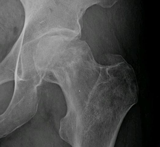
X-rays will usually show AVN if it has been present for long enough. In the very early stages, it may not show up on X-rays even though you are having pain. In the advanced stages, the hip joint will be very arthritic, and it may be hard to tell whether the main problem is AVN or advanced osteoarthritis of the hip. Either way, the treatment is basically the same.
If the X-rays fail to show AVN, you may have a bone scan done to determine if the pain in your hip is coming from early AVN. A bone scaninvolves injecting tracers into your blood stream. Several hours later, a large camera is used to take a picture of the bone around the hip joint. If there is no blood supply to the femoral head, the picture will show a blank spot where the femoral head should be outlined on the film.
The bone scan has pretty much been replaced with magnetic resonance imaging (MRI) today. The MRI scan is probably the most common test used to look for AVN of the hip. The MRI scanner uses magnetic waves instead of radiation. Multiple pictures of the hip bones are taken by the MRI scanner. The images look like slices of the bones. The MRI scan is very sensitive and can show even small areas of damage to the blood supply of the hip, even just hours after the damage has occurred.
Prognosis
Prognosis of avascular necrosis of femoral head is poor. Eventually almost all patients develop subchondral collapse, following which progression to osteoarthritis is inevitable.
This means that the dead bone in the head of femur becomes weak and breaks down. This causes the the hip joint to become incongruous. This causes osteoarthritis of the hip joint.
Treatment for Avascular Necrosis of femoral head
Although rest and exercise can sometimes heal the affected portion of bone, surgery is usually needed.
In as many as 80% of patients with early disease, an operation called core decompression can spark regeneration of the bone. In this procedure, the surgeon drills out the damaged section of bone up to the head of the femur. This opens up channels for blood vessels to reach the diseased area and foster the production of new bone. Hip pain is relieved, and as many as 75% of patients avoid joint replacement later on.
Early in the disease, osteotomy has been used to redistribute weight and prevent collapse and deformation of the femoral head. In patients with large areas of dead bone, however, osteotomy may hinder bone healing. In those patients—as well as for those with osteoarthritis or pain unrelieved by other treatments—total hip replacement is most often the treatment of choice.
Rehabilitation role in Avascular Necrosis of femoral head
Read more about AVN of Hip on Web MD
Nonsurgical Physical Therapy Rehabilitation
While physical therapy cannot cure avascular necrosis, in some cases it can help slow the progression of the disease and decrease the associated pain. A physical therapist can teach the correct way to use the appropriate assistive device (such as a cane or walker) to decrease weight bearing on the joint . They can provide proper exercises to help increase the strength of the muscles around the affect area (which will also decrease the weight on the joint). They may also use modalities such as electrical stimulation, ultrasound, joint mobilization, and heat to attempt to increase bloody supply to the area and help decrease pain.
You may work with a physical therapist who will show you ways to safely move and stretch your hip. The goal is to keep your hip mobile and to avoid losing range of motion. Your therapist will also instruct you to use a walker or crutches. Keeping weight off your hip while you are standing or walking may help the bone to heal while protecting the femur from further damage.
After Surgery Physical Therapy Rehabilitation
After a simple drilling operation, you will probably use crutches for six weeks or so. The drill holes weaken the bone around the hip, making it possible to fracture the hip. Using crutches allows the bone to heal safely and reduce the risk that you may fracture your hip. Patients who have had bone and blood vessels grafted are required to limit how much weight they place on the hip for up to six months.
When you are safe in putting full weight through the leg, your doctor may have you work with a physical therapist to help regain hip range of motion and strength.
Patients who require artificial hip joint replacement follow a structured program of physical therapy beginning shortly after surgery.
Return from Avascular Necrosis of Femoral Head to Orthopedic Physical Therapy
Return from Avascular Necrosis of Femoral Head to home page
Recent Articles
|
Author's Pick
Rating: 4.4 Votes: 252 |


 About Prodyut Das (PT, DPT) Physical Therapist at SMC, New York, USA. Former PT Winner Regional Health, South Dakota, Former HOD Physiotherapy & Fitness center @ NIMT Hospital, Greater Noida. Former PT ISIC Hospital. DPT ( Univ of Montana), MPT (neuro), MIAP, cert. manual therapist, Medical Neuroscience (USA). Licensed Physical Therapist in NY and Texas, USA. |
Connect with me- |
© Copyright physiotherapy-treatment.com since 2009 |