Enhance your health with free online physiotherapy exercise lessons and videos about various disease and health condition
Jersey Finger- Rupture of the Terminal Phalangeal Flexor
Jersey Finger represents rupture of the Flexor Digitorum Profundus. This usually happens when an athlete grabbing an opponent's shirt undergoes forced extension of the DIP while it is flexed. For this reason, it is called "grasping jersey finger" or "rugby finger". The ring finger is involved more often.
The rupture of the FDP tendon from its insertion on the distal phalanx known as Jersey Finger is often misdiagnosed as a sprained or "jammed" finger, as there is no characteristic deformity associated with it.
The injury is typically caused by forceful passive extension while the flexor digitorum profundus muscle is contracting. A common example is in football when the flexed finger is caught in a jersey while the athlete is attempting to make a tackle; hence the term jersey finger or rugby finger.
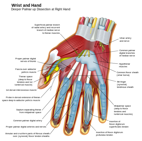 By The Photographer - Own work. Image renamed from Image:Wrist and hand deeper palmar dissection-en.svg, CC BY-SA 3.0, https://commons.wikimedia.org/w/index.php?curid=4433126
Sometimes the FDP disconnects from the base of 3rd phalanx, taking with it a small corticocancellous or bone fragment. This fragment can be seen in lateral x-ray of the finger, especially if enlarged, and can aid diagnosis. In other cases, the bone avulsion of flexor insertion is very large, so the 3rd phalanx undergoes traction of the extensor and tends to subluxate dorsally. The level of the retraction, the presence of a detached bone fragment, and age of the lesion allow the physician to suggest a classification of this type of lesion:
- 1. Type 1. The distal portion of the FPD withdraws until the crossing of the FDS and is sometimes blocked in this seat by a small bone fragment in front of the Phalanx I. The vincula longa is intact. In this most frequent type of lesion, especially if recent, the vascularization is retained and allows the tendon to be reinserted with a good chance of success. (The FDP tendon is torn but not ruptured)
- 2 . Type 2. The FDP is retracted (usually found in the palm). The vincula longa are broken and the whole extremity is devascularized. These forms are frequently seen in older lesions. (The FDP tendon is fully ruptured)
- 3. Type 3· There is a large epiphysial fragment of the Phalanx 3 that is blocked at the entrance of the digital canal. The DIP is destabilised in subluxations. (The FDP tendon ruptures and pulls a chip off the bone where it was attached)
Symptoms of jersey finger
- A pop or rip felt in the finger at the time of the injury
- Tenderness, swelling and warmth of the injured finger
- Pain when moving the injured finger and the inability to bend the last joint
- Occasionally a lump felt in the palm of the finger
- Bruising after 48 hours
Diagnosis
Physical Examination
To test the integrity of the tendon, the clinician isolates the FDP by holding the MCP and PIP joints of the affected finger in full extension, and asks the patient to attempt to flex the DIP. If it flexes, it is intact. If not, it is ruptured.
Plain radiograph
An x-ray to see if there is any damage to the bone.
Treatment
- Most mallet finger injuries at the DIP joint and most acute boutonnière deformities can be treated with splinting and the supervision of an occupational therapist.
- Use the physical examination to differentiate a boutonnière deformity from a pseudo boutonnière because the treatment is markedly different.
- Sagittal band rupture treated with splinting may take longer than with operative repair, and this may be a factor in the elite athlete.
- It is important to test for FDP function in any player who presents with DIP pain or stiffness. (Passively extend the PIP joint and observe for active FDP pull through at the DIP joint.)
- Conservative: for partial tear (i.e. splinting, NSAIDs, physical therapy)
- Surgical intervention: all complete flexor tendon injuries should be surgically repaired or at least referred to an orthopedic hand surgeon. Tendon retraction and time from injury are key. The tendon must be reattached to the distal phalanx. The rehabilitation of the condition is important as flexor tendons often become very stiff if not treated properly.
Physical Therapy for Jersey Finger
Because early motion of the healing finger is critical, passive flexion exercises begin about one week after surgery. These exercises, along with strengthening exercises using putty and hand therapy balls, can alleviate stiffness and reduced range of motion, frequent complications of finger surgery. Care must be taken not to introduce too much stress too early, or healing will be slowed. Physical therapy can also reduce the risk of repeated injury. Therapists perform wound care to the surgical incisions and move the fingers according to a specific protocol that will help restore finger range of motion, as well as pinch and grip strength to your finger.
Although most people recover full hand function following a jersey finger injury, they typically cannot return to playing sports for four to six months. In the meantime, regaining the use of fingers will enhance the quality of life and allow patient to perform daily activities free of pain and with maximum flexibility.
Read more about finger injuries & deformities


References
- DeLee & Drez's Orthopaedic Sports Medicine: Principles and Practicies
- G. Puddu· A. Giombini . A. Selvanetti (Eds.)-Rehabilitation of Sports Injuries
- Jersey Finger- Wikipedia
Back to home Page
Back to Sports physical therapy
Recent Articles
|
Author's Pick
Rating: 4.4 Votes: 252 |


 About Prodyut Das (PT, DPT) Physical Therapist at SMC, New York, USA. Former PT Winner Regional Health, South Dakota, Former HOD Physiotherapy & Fitness center @ NIMT Hospital, Greater Noida. Former PT ISIC Hospital. DPT ( Univ of Montana), MPT (neuro), MIAP, cert. manual therapist, Medical Neuroscience (USA). Licensed Physical Therapist in NY and Texas, USA. |
Connect with me- |
© Copyright physiotherapy-treatment.com since 2009 |