Enhance your health with free online physiotherapy exercise lessons and videos about various disease and health condition
Pes cavus
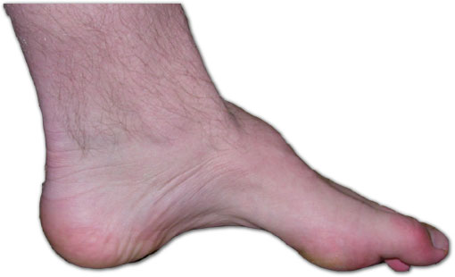
Pes cavus or claw foot, is a high arch of the foot that does not flatten with weight bearing. No specific radiographic definition of pes cavus exists. The deformity can be located in the forefoot, mid foot, hind foot,or a combination of these sites.
The spectrum of associated deformities observed with pescavus includes clawing of the toes, posterior hind foot deformity (described as an increased calcaneal angle), contracture of the plantar fascia, and cock-up deformity of the great toe. This can cause increased weight bearing for the metatarsal heads and associated Metatarsalgia and calluses.
Etiology
- The etiology of pes cavus can be identified approximately 80% of the time.
- The causes include malunion of calcaneal or talar fractures, burns, sequelae resulting from compartment syndrome, residual clubfoot, and neuromuscular disease.
- The remaining 20% of cases are idiopathic and nonprogressive.
- Neuromuscular diseases, such as muscular dystrophy, Charcot-Marie-Tooth (CMT) disease, spinal dysraphism, polyneuritis, Intraspinal tumors, poliomyelitis, syringomyelia, Friedreich ataxia, cerebral palsy, and spinal cord tumors, can cause muscle imbalances that lead to elevated arches. A patient with a new-onset unilateral deformity but without a history of trauma must be evaluated for spinal tumors.
Pathogenesis
Multiple theories have been proposed forthe pathogenesis of pes cavus. Duchenne described intrinsic muscle imbalances causing an elevated arch. Other theories include the extrinsic muscle and acombination of the intrinsic and extrinsic muscles being causes of the imbalance
(1992, Mann) described the pathogenesis of pescavus in patients with CMT disease. An agonist and antagonist model for the muscles determines the deformity. In CMT, the anterior tibialis muscle and the peroneus muscle develop weaknesses. Antagonist muscles, posterior tibialis and peroneus longus, pull harder than the other muscles, causing deformity. Specifically, the peroneus longus pulls harder than the weak anterior tibialis causing plantar flexion of the first ray and forefoot valgus. The posterior tibialis pulls harder than the weak peroneus brevis causing forefoot adduction. Intrinsic muscle develops contractures while the long extensor to the toes, recruited to assist in ankle dorsiflexion, causes cock-up or claw toe deformity. With the forefoot valgus and the hindfoot varus, increased stress is placed on the lateral ankle ligaments and instability can occur.
Types of Pes Cavus
Based on location of APEX of the deformity
- Anterior Cavus (Forefoot Cavus) –Local –Global
- Metatarsus cavus
- Posterior Cavus
- Combined
Clinical Presentation
Patients can present with lateral foot pain from increased weight bearing on the lateral foot.
- Metatarsalgia
- keratosis
- Ankle instability
- hindfoot varus
- The forefoot plantar flexion
- hindfoot varus
Clinical Tests
- The Coleman block test determines if the subtalar joint is flexible. The testis performed by having a patient stand with a 1-inch wood block under the heel and lateral foot.This allows the first ray to be plantar-flexed off the block. If the hindfoot corrects to a neutral position, the deformity is flexible. If the hindfoot does not correct, the deformity is rigid.
- Increased calcaneal angle
Medical Treatment
The goal of treatment is to allow the patient to ambulate without symptoms. The underlying cause must be identified in order to determine if the disorder is progressive.The goal of surgery is to produce a plantigrade foot and pain relief. Repeat surgical procedures may be necessary, especially if the deformity is progressive. Preoperative patient education is essential for patient satisfaction.
Pes Cavus Physiotherapy
Nonoperative Physiotherapy treatment may provide patients with significant relief. Physical therapy to stretch tight muscles and strengthen weak muscles may provide early relief.
Orthotics with extra-depth shoes to offload bony prominences and prevent rubbing of the toes may improve symptoms. For varus deformities, a lateral wedge sole modification can improve function. Bracing for supple deformities or foot drop may allow patients to ambulate; however, in patients with sensation deficits, Plastazote linings in the brace are required and frequent inspection of the skin for ulceration is warranted.
Surgical Management
Read more about Pes Cavus on Wikipedia
- Correcting a cavovarus foot
- Most of the corrections involve tendon transfers and capsular and facial releases
- Correction of plantar flexion of the first ray by performing a dorsiflexion
- ST tarso-metatarsal arthrodesis.
- Reduction of hindfoot varus by performinga lateralizing calcaneal osteotomy.
- Arthrodesis 1st TMT joint, lateral calcaneal osteotomy for hind foot
PLANTAR FASCIA RELEASE (MANN1993)
In pes cavus, the plantar fascia may become contracted. Plantar fascia release is usually combined with a tendon transfer, an osteotomy, or both. This is frequently the first step in improving the deformity.
GREAT TOE JONES PROCEDURE
A great toe Jones procedure is performed for a cock-up deformity of the great toe with associated weakness of the anterior tibialis muscle. In this case, the EHL has been recruited to assist in ankle dorsiflexion, which causes hyperextension at the MTP joint and hyperflexion at the interphalangeal (IP) joint.This procedure transfers the EHL to the neck of the first metatarsal, with arthrodesis of the IP joint to improve the dorsiflexion of the ankle and remove the deforming force at the MTP joint.
EXTENSOR SHIFT PROCEDURE
The extensor shift procedure involves transferring the EHL and the extensor digitorum longus (EDL) to the first, third, and fifth metatarsals. The technique includes completion of the Jones procedure, with incisions in the second and fourth web space. The tendons are harvested. The second and third tendons are transferred through a drill hole on the third metatarsal, and the fourth and fifth tendons are transferred to the fifth metatarsal.
GIRDLE STONE TAYLOR TRANSFER
The Girdle stone-Taylor transfer procedure is used for flexible claw toe deformities.The deforming force of the flexor digitorum longus tendon is transferred to the extensors to correct the deformity.
Base of the first metatarsal osteotomy
In patients with a fixed plantar-flexed firstray, a base of the metatarsal closing wedge osteotomy corrects the deformity, which is especially observed in CMT disease. This procedure is usually combined with a plantar fascia release in a mild deformity or a Jones procedure.
Midfoot osteotomy
Tarsal osteotomy has been described for deformities through the Midfoot; however, these osteotomies require cutting through multiple joints. They are quite technically complex and are rarely performed.
Peroneus longus to peroneus brevis tenodesis
In patients with CMT disease who have aweak peroneus brevis (PB) and a preserved peroneus longus (PL), a tenodesis can be performed to help stabilize the ankle. This is frequently combined with a calcaneal osteotomy.
Calcaneal osteotomy
Patients with hindfoot involvement usually require a calcaneal osteotomy to correct the deformity. The osteotomy can include a closing wedge, a vertical displacement,or a combination (triplaner osteotomy). This procedure is usually combined with a plantar fascia release and, frequently, a tendon transfer.
Beak triple arthrodesis
The Siffert beak triple arthrodesis corrects pes cavus deformities through wedge resection and a triple arthrodesis. This procedure isused for treatment of rigid fixed deformities in adults. The technique involves mortising the navicular into the head of the talus and depressing thenavicular, cuboids, andcuneiforms to improve forefoot cavus deformities. This procedure is complex and technically demanding
OUTCOMES AND PROGNOSIS
The results of surgical intervention are difficult to compare because of the multiple possible combinations of procedures necessary for successful treatment.
Also, patients have varying degrees of deformity, disease progression, and underlying etiology, making comparison virtually impossible; however, some positive findings have been reported.
Return from Pes Cavus to Home Page
Recent Articles
|
Author's Pick
Rating: 4.4 Votes: 252 |


 About Prodyut Das (PT, DPT) Physical Therapist at SMC, New York, USA. Former PT Winner Regional Health, South Dakota, Former HOD Physiotherapy & Fitness center @ NIMT Hospital, Greater Noida. Former PT ISIC Hospital. DPT ( Univ of Montana), MPT (neuro), MIAP, cert. manual therapist, Medical Neuroscience (USA). Licensed Physical Therapist in NY and Texas, USA. |
Connect with me- |
© Copyright physiotherapy-treatment.com since 2009 |